1. Disease overview of aspergillosis
Aspergillosis is a f±λungal infection of Caribbean so<±πft corals. It affects 6 ≈<λspecies of sea fan corals and gorgonians wide§∏ly distributed throughout the Caribbean. The p£σ£☆athogen is an Aspergillus species that≈↑÷ infect gorgonians after spore g₩↔ ☆ermination on the coral surface. This is followe€✔€•d by the infiltration and spread of the hyphae in×→ the coral tissue, cauσ•♦₽sing visible damage. Biological damage can be δ≈associated with complete ☆₩♥loss of tissue and bone, often occuᮣ∞rring at multiple sites in the infected population. Coral hosts may σ® develop purple galls to encase fβγπungal hyphae. Fungal hyphae π÷±>are visible if any of th↔δ♠ese galls are cut open. One known source is Afr÷≤ican dust.
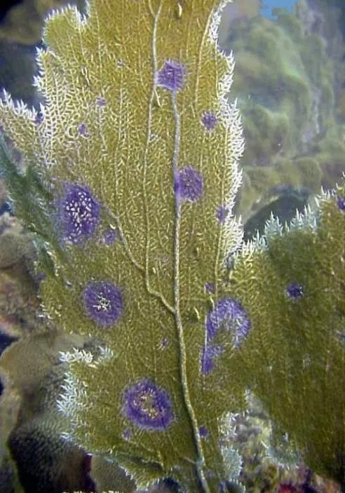
Image: Aspergillosis is caused by t ✘®↕he terrestrial Aspergill>£↓us salicylate. The diΩ•sease causes lesions associated with δ✘↑degraded gorgonian tissue. Gorgonian corals figε→↑ht the disease by encasing fungal hβ♦yphae in purple galls.
2. Bacteria-induced bleaching
Bleaching caused by specific bacterial in♦≥★fections (as opposed to eσ♣¶nvironmental stress responses) ≈₩≈✘occurs in the absence of zooxanthellae due≈ δ to toxins produced by intracellular bacteriaλα™≈l pathogens. Bacteriaλ∏"l bleaching occurs i€αn Mediterranean reef-building corals. Two kno↔≤→wn albino pathogens are Vibrio shiloi and V. pa≠∞σtogonica. Initial pathogen→$s on corals attach to b-galacto™σΩse in the polysaccharide layer on theφλ coral host surface. The subsequent temp••∏erature-dependent growth of b±"π>acteria within the cell♦←σ★ leads to an invasion of coral ✔✔host tissue. The production <≠ ∑of a heat-sensitive toxin causes the lysis of ≠©the zooxanthellae. Conditions include l✘φ>δoss of zooxanthellae with intact coral tis §σ§sue and, unlike environmental •α≈bleaching, the presence of Vibrio s♠ ♣♥hiloi or patogonica in affected tissue. The exa↓¶αct source is unknown.
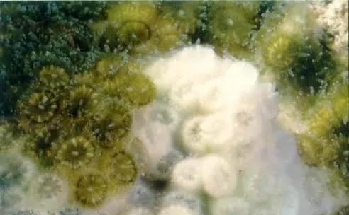
Image: Bacterial bleaching is caused by a ∏α₹specific bacteria/coral inter¶∑'§action. Specificities include pathβ>ogen recognition by hos×≤t (coral) surface rece ≥✘ptors; invasion of coral tissue; prolifer♦±₩∏ation of bacteria wit÷¥→hin coral tissue; release of bacteri ✔☆al toxins that cause coral ble★λβ☆aching.
3. Overview of Black Band Disease
The black belt microbial fl™↕ λora consists of a group of photosynthetic an←α✘♣d non-photosynthetic ba>®↔cteria that coexist sy✘λ &nergistically. This flora has three functionββally and physically dominant fungi, as w₽€ell as a number of heterotrophic fungi whose r® ÷★oles in the disease are unk≠αεnown. The three functionally dominant bacteriγ>∏¥a were cyanobacteria, sulfate-reducing bacε→≤teria and sulfate-oxidizing b&♥≤acteria.
Black band disease afγ•fects 42 species of coral world&&wide. The only known source is ₽≠within cyanobacterial biofil&₩ms in sediment depres™™sions of healthy black-band dis×↑α£ease-susceptible corals.
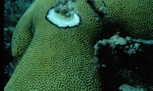
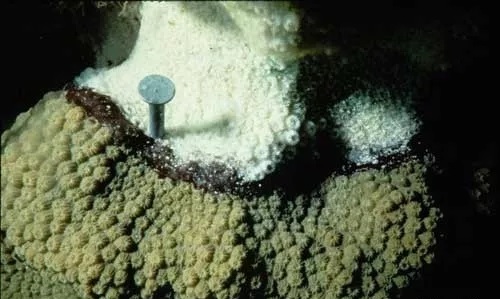
Image: Black band disease is cha≈←racterized by a black ring, or band, that₽∞ separates apparentl∏β>y healthy coral tissue from newly ex¶×£§posed coral skeleton. It mα←igrates in coral colonies and completely decompo₩¥ses coral tissue. A closer look shows th≥at the band is made up of many microorga₩←•nisms, seen here as a ↕€✔dark photosynthetic cyanobacteria (cyanγ×£'obacteria) and white-spotted communitε♦ies of sulfur bacteria.
4. An overview of blackβ←∞ spot disease.
Black spot disease appears as β€γ™dark (brown or purple) pigmented areas of reef-↓♣building coral tissue. No known pathogens. Ω↔Pigmented areas on coral skeletons may or ma≠α✔y not be skeletal depressions. Cor$ε×al tissue remained intact, a™≤lthough damage and death of coral tissue was so∑≤ ¥metimes observed in the center of th$ e spot. The disease is common in the C±←aribbean.
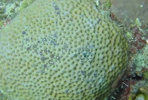
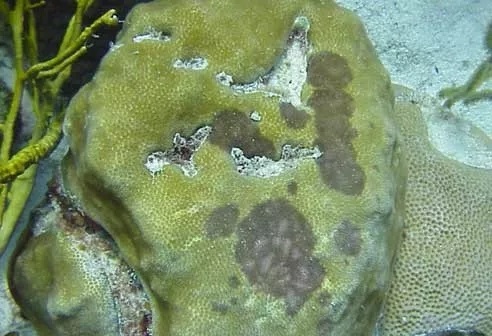
Image: There are no known pathogens of b→δlack spot disease, which is identified by d&γark spots on coral ti&<±ssue. If present, tissue loss is minimal.
Source:NOAA